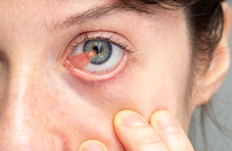


A pterygium is a growth which arises on the conjunctiva and grows towards and infiltrates, the surface of the cornea. As it grows, it typically forms a triangle with the head of the pterygium towards the center, and the body and tail (the base of the triangle) towards the canthus (the point where the upper and lower eyelids meet). The elevated growth typically develops over the edge of the cornea and grows inward where it eventually may cause impaired vision.
The origin of pterygia is not completely clear and doctors are still researching the exact cause of a pterygium. Studies show high exposure to ultraviolet light and dry conditions increase the probability of the growth. Pterygia are more common for patients in warm climate areas. Trauma from exposure to pollen, dust, sand, wind, smoke and other environmental stimuli can also add to the risk of pterygium formation. If sunglasses are not worn in the regions of sunny climates, this increases the risk of developing pterygia. People with light color eyes and light skin pigmentation may also have a higher risk, too.
The symptoms of pterygium are often not severe, but may include eye irritation, redness, and blurred vision. Most patients complain of itchiness, scratchiness and burning. A pterygium grows slowly, and may not affect your vision unless it grows directly over the center of the cornea. A pterygium can also cause contact lens intolerance.
There are a number of things that can be confused with a pterygium and may require a biopsy.
Pseudo-pterygium is the result of a healing response from the body to an external injury. When the eye develops an inflammatory conditions from a corneal ulcer (infection) or inflammation from chemical, auto immune or microbiological insults, the conjunctiva mobilizes itself to heal the area of damage. This can also happen as a result of healing from pterygium surgery. A pseudo-pterygium tends to be stationary.
Pterygiums can have variations in growth patterns, as they can stop growing or undergo sudden reactivation of growth. Pterygiums usually grow slowly, but their growth can be unpredictable. Some pterygia can stop growing after a long period of growth.
Pterygiums have a stationary (inactive) phase and a more advanced rapid growing phase. Clinical examination can sometimes tell you if your pterygium is active. A Stocker line, a fine line representing iron deposition in the cornea and in front of the leading edge of the pterygium, is a sign the pterygium is probably in the stationary phase. Rapidly growing pterygiums do not allow enough time for the iron to deposit. In active pterygiums, the body of the pterygium has of vessels versus a whitish look to a stationary one. Epithelial microulcerations that stain with fluorescein can be seen during an exam in active pterygiums. Pterygiums can become red or irritated, in that case, your doctor may prescribe eye drops and ointments to relieve inflammation and irritation. Not all pterygiums require surgery.
Surgical treatment for Pterygium is not necessary unless the pterygium is irritating despite the use of artificial tears, is causing astigmatism or visual loss, or is approaching the line of vision. In many instances, patients prefer to have the pterygium removed for cosmetic purposes. You should be aware that pterygia can grow back after surgery rapidly and sometimes, violently. Patients also experience dryness and irritation after the removal, but surface lubrication and other medications can be used as treatment and to help prevent recurrence.
Many alternatives have been suggested for the surgical treatment of pterygiums. During bare sclera excision, an area of the white part of the eye is left uncovered as a barrier for growth into the cornea. In primary closure, the pterygium is removed and the conjunctiva closed. Conjunctival autografting or the removal and transplantation of healthy conjunctiva not exposed to the sun from a different part of the eye, results in a lower rate of recurrence than primary closure. However, sutures are used to secure the tissue to its new location. This increases the amount of postoperative discomfort and may increase the amount of inflammation. Alternatively, amniotic membrane grafts can be used. During this technique, instead of harvesting the conjunctiva, a piece of donor amniotic membrane is glued in place using TISSEEL Duo Quick (Baxter). The use of glue instead of sutures leads to less postoperative pain, faster recovery and shortened surgical time. The rate of recurrences are similar between conjunctival autografts and amniotic membrane grafts which are a lot lower than primary closure. Some research studies have suggested higher rates of recurrence with autografts and others with amniotic membrane grafts.
Every surgery has some risks but, pterygium surgery is usually tolerated well and has a very low rate of complications. Every patient will experience some swelling, bruising, tearing, sensitivity to light and some discomfort. There is always the risk of bleeding, infection double vision due to scarring, or muscle damage although highly unlikely. The cosmetic appearance after pterygium is usually good, but usually not perfect as there can be scarring and vessel growth. Our doctors will discuss the pertinent risks before surgery. A theoretical risk never reported is transmission of parvovirus B19, hepatitis or human immunodeficiency virus from fibrin glue. This glue has been used in vascular and abdominal surgery for almost a decade without any reported cases or complications specific to the glue.
Mitomycin C is a chemotherapy drug isolated from the fermentation filtrate of some Streptomyces bacterial species; it can generate free radicals that lead to tissue breakdown and also inhibit DNA, RNA and protein synthesis.
After pterygium removal, cells called fibroblasts begin to heal the area and can cause recurrences. MMC has been used to eliminate these fibroblasts and decrease the rate of recurrence after pterygium surgery. MMC has been used before surgery to shrink the pterygium, during and after surgery. MMC, however, is not without risks, as there have been reports of corneal and scleral melts and perforation. In most cases, ophthalmologists will use MMC in low concentrations (0.02% for short) applications of 3 minutes; the likelihood of complications with this dosage of MMC, is low.